)
It's mid-morning, and you've just finished an abdominal scan on a vomiting Labrador. The liver looked mildly hyperechoic, the spleen had a small nodule worth noting, and you took measurements on both. You move straight into the next appointment.
By the time you sit down to write the referral note, some of that detail has already started to blur. Was the splenic nodule 0.8 cm or 1.2 cm? Did you note the gallbladder wall thickness? Your practice doesn't have a standard format for these reports, and last week, two different clinicians documented the same type of case in two different ways.
According to the American College of Veterinary Radiology and European College of Veterinary Diagnostic Imaging, Veterinary imaging reports are official medicolegal documents that serve as the primary means of communication for radiologists and significantly influence clinical decisions. A structured ultrasound report template helps make sure that what happened during the scan and what ends up in the record are the same thing.
This article covers what a complete ultrasound report should include, how POCUS reports differ from comprehensive studies, and how a standardized template supports consistent documentation across your team.
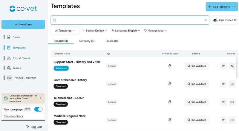
CoVet has a growing database of veterinary templates. Check out other templates that might be helpful for you:
What a veterinary ultrasound report template is and when you need one
A veterinary ultrasound report template provides a consistent structure for documenting a sonographic exam: what was scanned, what was seen, what it likely means, and what should happen next.
The template stays the same regardless of the case, but the content inside it adapts to the patient, the clinical question, and the type of scan performed.
Comprehensive abdominal ultrasound vs. POCUS: what the report needs to capture
Scope | Who performs it | Documentation depth | When it's used |
Comprehensive abdominal ultrasound | General practitioner, sonographer, or board-certified radiologist | Full organ survey, standardized measurements, detailed impression | Referrals, diagnostic workup, pre-surgical planning |
Point-of-care ultrasound (POCUS) | Attending clinician, often in real time | Focused on a specific clinical question, views obtained, findings per view | Emergency triage, monitoring, procedural guidance |
A structured veterinary report template helps ensure findings, impressions, and recommendations are communicated consistently, whether the exam is a full abdominal ultrasound or a focused POCUS scan.
When a written report is required
A written ultrasound report generally matters most when:
The patient is being referred to a specialist
The exam is part of a telemedicine consult
A board-certified radiologist is reviewing images remotely
The findings need to be communicated to a referring veterinarian
The case could become part of a medicolegal review later
For practices working with telemedicine or remote interpretation, veterinary radiology workflows often add an additional layer of documentation expectations, since the interpreting radiologist may never see the patient directly.
The ACVR and ECVDI consensus statement notes that imaging reports hold legal significance as evidence of patient care and serve as the main method of communication for radiologists, which is part of why standardized formatting matters even for routine cases.
A consistent veterinary ultrasound report template helps ensure that standard is met whether the report is written manually or generated through an AI scribe like CoVet.
What is included in CoVet's veterinary ultrasound report template
CoVet's ultrasound report template covers five core areas: patient and study information, clinical history, sonographic findings, interpretation and assessment, and recommendations.
Patient and study information
Patient ID
Species, breed, age, sex
Body weight
Date of examination
Referring veterinarian
Equipment used
Operator credentials
A standardized patient history form completed at intake can help ensure this section is already populated when the imaging report is started.
Clinical history and indication
The clinical history section explains why the patient is being scanned.
This isn't a formality: research has shown that the same imaging finding can lead to different interpretations across patients, since radiologists often lack complete patient information and their impressions are based on the available data.
A finding like mild hepatomegaly means something different in a vomiting six-month-old puppy than in an asymptomatic twelve-year-old dog on long-term medication.
CoVet's template captures this clinical history before the findings section, and includes:
Presenting complaint
Relevant medical history
Current medications
Reason for imaging (the specific clinical question)
Interpretation and assessment
Findings are objective observations: what was seen, measured, and described.
Impression, on the other hand, is the clinical interpretation: what those findings likely mean, given the patient's history and presentation.
CoVet's template keeps these as separate sections, mirroring the assessment portion of a veterinary SOAP note: a space for clinical reasoning, not just a repeat of the observations.
Recommendations
The recommendations section translates the impression into next steps:
Further diagnostics (bloodwork, additional imaging, biopsy)
Follow-up imaging timeline
Referral to a specialist
Monitoring plans
Repeat examination interval
When recommendations include instructions the client needs to follow at home, a discharge instructions template keeps that information separate from the clinical report while ensuring it still gets communicated.
Automating your veterinary ultrasound report template
CoVet is an AI-powered veterinary scribe and copilot. It uses natural language processing to turn what you say during or after a consultation into structured clinical notes — including ultrasound reports formatted by organ system, with findings, interpretation, and recommendations already in place.
It produces a structured, actionable clinical record in approximately 30 seconds. The ultrasound report template is one of 95+ templates built by practicing veterinarians, covering everything from SOAP notes to specialty workflows.
Records can be exported to supported PMS systems or transferred using the Chrome extension.
This kind of structured documentation also matters for where veterinary imaging is heading. CoVet also works offline, which supports field and mobile veterinary work where connectivity isn't reliable.
Frequently asked questions
Is a POCUS report the same as a full abdominal ultrasound report?
No. A POCUS report is focused and question-driven: it documents the specific clinical question being asked (such as "is there free fluid?") and the views obtained to answer it.
What's the difference between findings and impressions in an ultrasound report?
Findings describe what was directly observed during the scan: organ size, echogenicity, presence of fluid, and so on. The impression is the clinical interpretation of those findings, taking into account the patient's history and presentation. Keeping these sections separate makes it easier for the reader to distinguish between raw observations and clinical reasoning.
How do I document a normal ultrasound?
Normal findings still need to be documented organ by organ, not summarized as "no significant abnormalities."
A complete record of what was evaluated and found to be within normal limits is part of the medicolegal value of the report, and it gives future clinicians a baseline for comparison if the patient is scanned again later.
Does my ultrasound report need to include measurements?
For most organs, yes. Standardized measurements provide an objective baseline that supports the impression and allows for comparison on follow-up exams.
While specific requirements can vary, guidance from groups like the ACVR and ECVDI generally points toward including measurements for organs where size or thickness is clinically relevant, such as the liver, spleen, kidneys, and bladder wall.
Can I use the same template for dogs and cats?
The overall report structure works for both species, since the core sections (patient information, clinical history, findings, interpretation, recommendations) don't change.
What does change is the normal reference ranges for organ measurements and some species-specific considerations in interpretation. Species-specific details captured during intake on a patient history form can help make sure the right context carries through into the imaging report.
About the Author
)
)
)